
High Tibial Osteotomy (Leg Curvature Treatment)
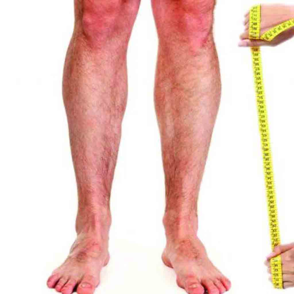
As the knee cartilage wears down, the shin bone (tibia) begins to angle inward relative to the body's weight-bearing axis. Because of this angulation, a greater share of the body's weight presses on the inner side of the knee, and the knee, caught in this vicious cycle, rapidly develops osteoarthritis. This cartilage damage (osteoarthritis) eventually leads to the need for a knee replacement.
The main goal of this surgery is to take the load off the worn inner side of the knee and transfer it to the healthier outer side.
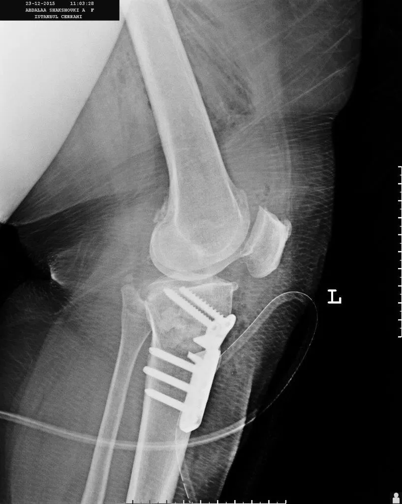
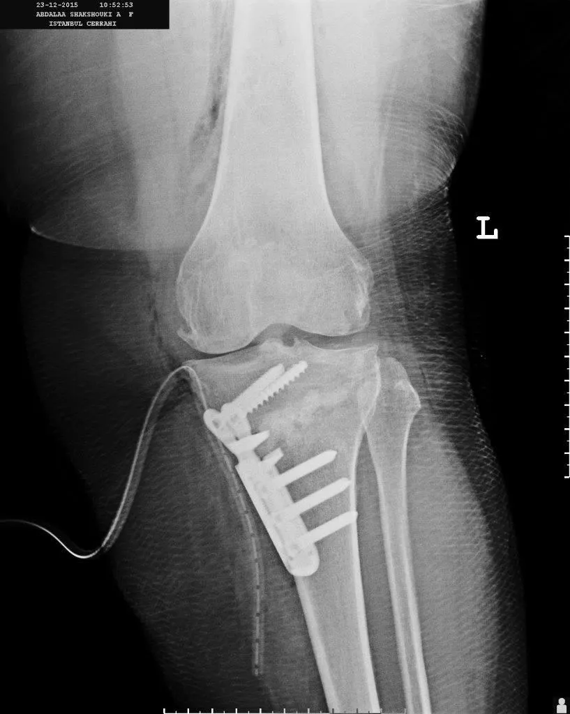
Who is high tibial osteotomy suitable for?
Classically, it is performed on patients younger than 60 who have osteoarthritis on the inner side of the knee but no advanced osteoarthritis on the outer side of the knee or in the kneecap.
On preoperative examination, there should be no significant restriction of motion in the knee joint, and it should be established that the symptoms originate from the inner part of the knee.
In addition, a full-length leg X-ray should show that the leg axis (mechanical axis) has shifted inward, and the correction should be planned according to the measured angle.
What are the advantages and disadvantages of high tibial osteotomy?
- When performed in the right patient, it allows osteoarthritis to be treated while preserving the patient's own joint.
- There is a high likelihood that it will spare the patient a knee replacement for life.
- There is no risk of revision (loosening), as there is with knee replacements.
- Whereas patients walk with full weight-bearing early after replacement surgery, after high tibial osteotomy it may take 4–6 weeks to progress to full weight-bearing, depending on the patient's weight, the amount of correction and the security of the fixation.
- If osteoarthritis develops after high tibial osteotomy and conversion to a knee replacement becomes necessary, the osteotomy does not make the replacement technique more difficult.
- After high tibial osteotomy, it is not possible to predict how much of the preoperative pain will be relieved, whereas pain is brought under control after replacement surgery.
High Tibial Osteotomy Surgical Technique
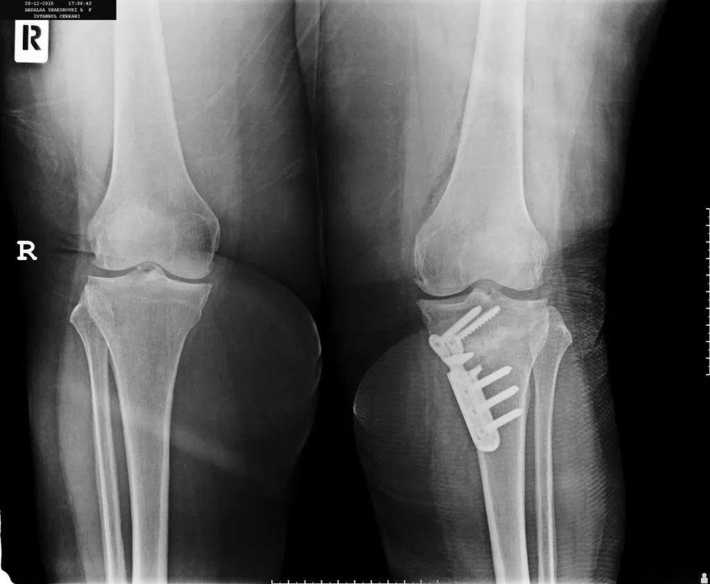
Before the open surgery, knee arthroscopy is performed to visualize the cartilage damage on the inner side of the knee and to examine the cartilage of the outer side of the knee and the kneecap. If all of these structures are suitable for high tibial osteotomy, the open surgery begins once the arthroscopic procedures (meniscectomy, cartilage debridement, etc.) are completed.
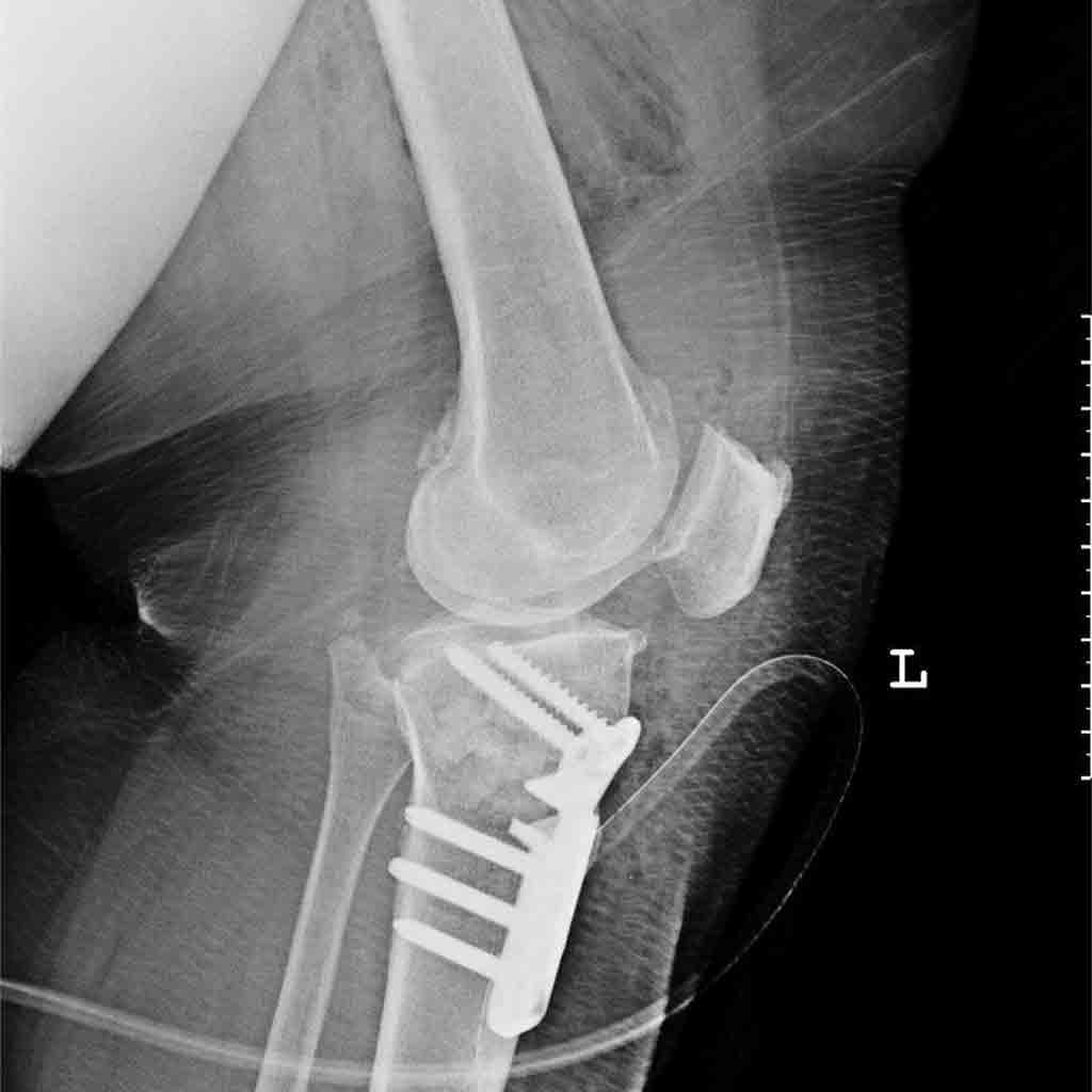
The tibia is approached from its inner side, close to the joint. After the bone surface is prepared, staying in front of the medial collateral ligament, 2 parallel guide K-wires are inserted under fluoroscopy, and an incomplete osteotomy (bone cut) is made just below these guide wires. The final part of the osteotomy is completed with osteotomes, stopping at least 1 cm short of both the outer cortex and the joint line, and the bone is then gradually levered open. The bone, opened to the required angle, is fixed with a plate and screws. Depending on the surgeon's preference, an autograft or allograft can be placed in the gap.
Common errors in this surgery include cutting through the outer part of the bone as well, or the outer part breaking while the bone is being opened; fracture lines extending into the joint; inadequate fixation; and opening the bone too far toward the front or back in the side (sagittal) plane, which alters the angle the joint makes with the ground (tibial slope).

The Postoperative Period
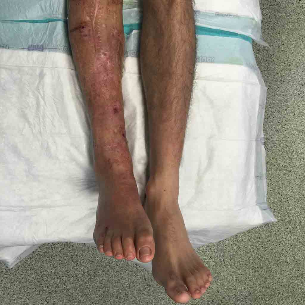
- When you come out of surgery, you will see that the inward curvature of your leg has been visibly corrected when you look at it.
- The drain (blood drainage tube) placed in your knee is removed after 24–48 hours.
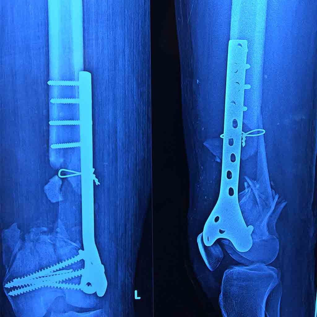
- During the visit in the first minutes after surgery, you will be informed about the details of the operation and any points specific to your case, using the X-ray taken in the operating room and photographs taken during the surgery.
- On the evening of the surgery or the following morning, a team made up of a physiotherapist, a nurse and myself will help you stand up and walk with a walker (four-legged walking frame) or crutches.
How much weight you can put on the leg is closely related to the surgical technique and your body weight. - To prevent venous thromboembolism (a blood clot forming in the leg) after surgery, protection is provided with low-molecular-weight heparin (Clexane®), ankle pumping exercises, anti-embolism stockings and early mobilization.
- Antibiotics are stopped once your drain has been removed.
- You will be discharged after approximately 2–3 days.
- After discharge, it is important to keep up the in-bed exercises you will do at home and your walking, to take painkillers if you have pain, and to continue the blood thinner given to you.
- Your stitches are the self-dissolving type, and you can let water run over your wound and wash from the 2nd week after surgery. Scrubbing the wound is not recommended; the suture ends will fall off on their own.
- A follow-up X-ray is taken during the first month after surgery.
- The implant placed in your knee does not need to be removed. Removal of the implant is planned only in special circumstances.